
STAMICIS KIT FOR RADIOPHARMACEUTICAL PREPARATION 1MG [SIN14129P]
Active ingredients: STAMICIS KIT FOR RADIOPHARMACEUTICAL PREPARATION 1MG
Last updated 21 July 2026
Product Info
STAMICIS KIT FOR RADIOPHARMACEUTICAL PREPARATION 1MG
[SIN14129P]
Product information
Active Ingredient and Strength | TETRAKIS (2-METHOXYISOBUTYL ISONITRILE) COPPER (I) TETRAFLUOROBORATE - 1 MG |
Dosage Form | INJECTION, POWDER, FOR SOLUTION |
Manufacturer and Country | CIS BIO INTERNATIONAL - FRANCE |
Registration Number | SIN14129P |
Licence Holder | QT INSTRUMENTS (S) PTE LTD |
Forensic Classification | PRESCRIPTION ONLY MEDICINES |
Anatomical Therapeutic Chemical (ATC) code | V09GA01 |
Prescription-only Medicines with Exemptions for Supply without Prescription | NA |
Indication
4.1. Therapeutic indications
This medicinal product is for diagnostic use only.
This is indicated for adults. For paediatric population see section 4.2.
After radiolabelling with sodium pertechnetate (99mTc) solution, the solution of technetium (99mTc) sestamibi obtained is indicated for:
Myocardial perfusion scintigraphy for the detection and localisation of coronary artery disease (angina pectoris and myocardial infarction).
Assessment of global ventricular function. First-pass technique for determination of ejection fraction and/or ECG-triggered, gated SPECT for evaluation of left ventricular ejection fraction, volumes and regional wall motion.
Scintimammography for the detection of suspected breast cancer when mammography is equivocal, inadequate or indeterminate.
Localisation of hyperfunctioning parathyroid tissue in patients with recurrent or persistent disease in both primary and secondary hyperparathyroidism, and in patients with primary hyperparathyroidism scheduled to undergo initial surgery of the parathyroid glands.
Dosing
4.2 Posology and method of administration
Posology
Adults and elderly population
Posology may vary depending on gamma camera characteristics and reconstruction modalities. The injection of activities greater than local DRLs (Diagnostic Reference Levels) should be justified.
The recommended activity range for intravenous administration to an adult patient of average weight (70 kg) is for:
Diagnosis of reduced coronary perfusion and myocardial infarction
The recommended activity range for diagnosis of ischaemic heart disease according to the European Nuclear Medicine Guide of the EANM 2019 (European Association of Nuclear Medicine) is:
– Two-day protocol: 300–600 MBq/study if a conventional Anger gamma camera is used, or 180–500 MBq if a cardiac-centered gamma camera is used for acquisition
– One-day protocol: 250–400 MBq for the first injection, three times more for the second injection if a conventional Anger gamma camera is used, or 150–300 MBq if a cardiac-centered gamma camera is used for acquisition.
Not more than a total of 1,600 MBq should be administered for a one-day protocol and 1,200 MBq for a two-day-protocol. For a one-day protocol, the two injections (stress and rest) should be done at least two hours apart but may be performed in either order. After the stress injection, exercise should be encouraged for an additional one minute (if possible).
For diagnosis of myocardial infarction one injection at rest is usually sufficient.
For diagnosis of ischaemic heart disease two injections (stress and rest) are required in order to differentiate transiently from persistently reduced myocardial uptake.
Assessment of global ventricular function: 600–800 MBq injected as a bolus.
Scintimammography: 700–1000 MBq injected as a bolus usually in the arm opposite to the lesion.
Localisation of hyperfunctioning parathyroid tissue:
For the dual phase technique 400–900 MBq injected as a bolus. The typical activity is between 500–700 MBq.
For the subtraction technique with sodium pertechnetate (99mTc): 75–110 MBq sodium pertechnetate (99mTc) by intravenous administration followed by 400–900 MBq (99mTc) sestamibi by intravenous administration, or 400–900 MBq (99mTc) sestamibi by intravenous administration followed by 150 MBq sodium pertechnetate (99mTc) by intravenous administration.
For the subtraction technique with sodium iodide (123I): 7.5–15 MBq sodium iodide (123I) administered orally or intravenously followed 2 hours later by 400–900 MBq (99mTc) sestamibi by intravenous administration.
Renal impairment
Careful consideration of the activity to be administered is required since an increased radiation exposure is possible in these patients.
Hepatic impairment
In general, activity selection for patients with a decreased hepatic function should be cautious, usually starting at the low end of the dosing range.
Paediatric population
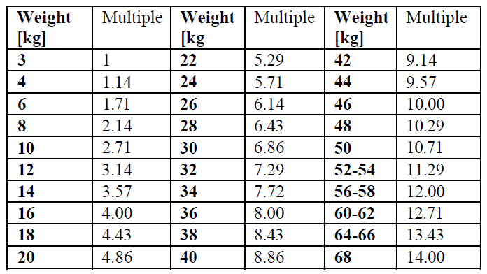
The use in children and adolescents has to be considered carefully, based upon clinical needs and assessing the risk/benefit ratio in this patient group. The activities to be administered to children and adolescents may be calculated according to the recommendations of the European Association of Nuclear Medicine (EANM) paediatric dosage card; the activity administered to children and to adolescents may be calculated by multiplying a baseline activity (for calculation purposes) by the weight-dependent multiples given in the table below.
A[MBq]Administered = Baseline Activity X Multiple:
The baseline activity is 63 MBq as a cancer seeking agent. For cardiac imaging, the minimum and maximum baseline activities are 42 and 63 MBq, respectively, for the two-day protocol cardiac scan both at rest and stress. For the one-day cardiac imaging protocol, the baseline activity is 28 MBq at rest and 84 MBq at stress. The minimum activity for any imaging study is 80 MBq.
Method of administration
For intravenous use.
Because of potential tissue damage, extravasal injection of this radioactive product has to be strictly avoided.
For multidose use.
Precautions to be taken before handling or administration of the medicinal product
This medicinal product should be reconstituted before administration to the patient. For instructions on reconstitution and control of the radiochemical purity of the medicinal product before administration, see section 12 – please refer to the Product Insert/Patient Information Leaflet published on HSA for the full drug information.
For patient preparation, see section 4.4 – please refer to the Product Insert/Patient Information Leaflet published on HSA for the full drug information.
Image acquisition
Cardiac imaging
Imaging should begin approximately after 30–60 min after injection to allow for hepatobiliary clearance. Longer delay can be required for resting images and for stress with vasodilators alone because of the risk of higher subdiaphragmatic technetium (99mTc) activity. There is no evidence for significant changes in myocardial tracer concentration or redistribution, therefore imaging for up to 6 hours post injection is possible. Test may be done in a one day or two days protocol.
Preferably tomographic imaging (SPECT) with or without ECG gating should be performed.
Scintimammography
Breast imaging is optimally initiated 5 to 10 minutes post injection with the patient in the prone position with breast freely pendant.
The product is administered in an arm vein contralateral to the breast with the suspected abnormality. If the disease is bilateral, the injection is ideally administered in a dorsal vein of the foot.
Conventional gamma camera
The patient should then be repositioned so that the contralateral breast is pendant and a lateral image of it should be obtained. An anterior supine image may then be obtained with the patient’s arms behind her head.
Camera dedicated to breast imaging
In case a camera dedicated to breast imaging is used, a relevant machine-specific protocol must be followed to obtain the best possible imaging performance.
Parathyroid imaging
Parathyroid image acquisition depends on the protocol chosen. The most used studies are either the subtraction and/or the dual-phase techniques, which can be performed together.
For the subtraction technique either sodium iodide (123I) or sodium pertechnetate (99mTc) can be used for imaging for the thyroid gland since these radiopharmaceuticals are trapped by functioning thyroid tissue. This image is subtracted from the technetium (99mTc) sestamibi image, and pathological hyperfunctioning parathyroid tissue remains visible after subtraction.
When sodium iodide (123I) is used, images are acquired simultaneously, starting 5 min after injection of (99mTc)sestamibi. Images are inspected visually, normalized to thyroid counts, and sodium iodide (123I) images are subtracted from the (99mTc)sestamibi images.
When sodium pertechnetate (99mTc) is used, image acquisition of sodium pertechnetate (99mTc) is starting 20–30 minutes after injection. Image acquisition of (99mTc)sestamibi is starting 10–15 minutes after injection. Images of sodium pertechnetate (99mTc) are either digitally or cognitively subtracted from the (99mTc) sestamibi images.
When the dual phase technique is used, the first neck and mediastinum image is obtained 10 minutes later. After a wash-out period of 1 to 2 hours, neck and mediastinum imaging is again performed.
The planar images may be complemented by early and delayed SPECT or SPECT/CT.
Contraindications
4.3 Contraindications
Hypersensitivity to the active substances to any of the excipients listed in section 6.1 – please refer to the Product Insert/Patient Information Leaflet published on HSA for the full drug information or to any of the components of the labelled radiopharmaceutical.
In myocardial scintigraphy investigations under stress conditions, the general contraindications associated with the induction of ergometric or pharmacological stress should be considered.